the blueprint of life [11]: DNA replication 1

DNA replication

Definition: the process of copying a parental DNA molecule to form two daughter DNA molecules.

  • Introduction to DNA replication
  1. DNA replication is essential for cell proliferation, i.e. mitosis, meiosis.
  2. DNA replication is a complex endeavor involving a series of enzyme activities.→see “DNA polymerases”
  3. DNA replication is performed in a semiconservative and semidiscontinuous mode.→see “DNA replication is semi-conservative”
  4. —DNA replication has 3 stages: initiation, elongation and termination.→next section
  5. —DNA replication is tightly regulated, involving various protein-protein, protein-DNA interactions.
  6. —DNA replication of prokaryote and eukaryote shares similar features, but is distinctive in details.
  •  Chemical Reaction of DNA replication

Essentials

1. Substrate: deoxynucleoside triphosphates(dNTPs)
2. Template: a primer-template junction
       DNA is synthesized by extending the 3’ end of the primer (free 3’-OH is required)
       – RNA primer or priming from a nick in DNA
3. Enzymes: DNA polymerases etc
4. Energy supply: Hydrolysis of pyrophosphate (PPi) is the driving force for DNA synthesis
5. Ions involved: Mg++ or Zn++
  • DNA polymerases
  • DNA polymerase I—
    • Pol I was the first enzyme discovered with polymerase activity, and it is also the best characterized one.
    • —Although abundant in cells (400/cell), Pol I is NOT the primary enzyme involved with bacterial DNA replication.
    • Main functions of Pol I: — (1) Fill any gaps in the new DNA that result from the removal of the RNA primer by its 5’ -3’ polymerase activity;   (2) Remove a new mispaired base by proofreading (校读)3’-5’ exonuclease (外切酶) activity. (3)Remove the RNA primer by its 5’-3’ exonuclease activity
The 3′–>5′ exonuclease activity intrinsic to several DNA polymerases plays a primary role in genetic stability; it acts as a first line of defense in correcting DNA polymerase errors. A mismatched basepair at the primer terminus is the preferred substrate for the exonuclease activity over a correct basepair. (source)
  • DNA polymerase III
    • The primary polymerase in DNA replication, although lower in abundance (15/cell)than pol  →referred to as “replicase
    • functions: (1) 5’→3’ polymerase activity; (2) 3’→5’ exonuclease activity – proofreading
    • Catalytic efficiency: much higher than pol I→High processivity and polymerization rate
    • A multi-unit complex: “holoenzyme” (全酶)
  • DNA replication is semi-conservative 

Bet you all have learned it in high school, and the famous experiment by Meselson and Stahl. We still need to go over the points again as they are essentially important for what we will learn next.

The key to the mechanism of DNA replication is the fact that each strand of the DNA double helix carries the same information-their base sequences are complementary (we talked about this in THE BLUEPRINT OF LIFE [2]: PRIMARY AND SECONDARY STRUCTURE OF DNA).

During replication, the two parental strands separate and each acts as a template (that’s right, the template for DNA replication is DNA itself!)to direct the enzyme-catalyzed synthesis of a new complementary daughter strand with the normal base-pairing rules (A-T, C-G)

This semi-conservative mechanism was demonstrated experimentally in 1958 by Meselson and Stahl.

Hypotheses:

In the experiment:

E. coli cells were grown for several generations in presence of the stable heavy isotope 15N so that their DNA became fully density labeled (both strands are 15N labeled: 15N/15N)

The cells were then transferred to medium containing only normal 14N and, after each cell division, DNA was prepared from a sample of the cells and analyzed on a CsCI gradient using the technique of equilibrium (isopycnic) density gradient centrifugation, which separates molecules according to differences in buoyant density.

After the first cell division, when the DNA had replicated once, it was all of hybrid density, in a position on the gradient half way between fully labled (15N /15N )and fully light (14N/14N). After the second generation in 14N, half of the DNA was hybrid density and half fully light.

Thus, two of the hypotheses were denied, left us with the semi-conservative mechanism.

After each subsequent generation, the proportion of 14N/14N increased, while some DNA of hybrid density persisted. Thus the semi-conservative mode of DNA replication is confirmed: each daughter molecule contains one parental strand and one newly-synthesized strand.

DNA replication & PCR, General Biology, Open Courses at UC-Berkeley

General Biology, Great open courses given by Gary L. Firestone, Michael Meighan Jasper D. Rine and Jennifer A. Doudna, professors at UC-Berkeley.

There is more to DNA replication than we talked about yesterday so I tried to upload this video to help you learn more. 

“tried? ”

“Well, turned out ‘ DNA replication and PCR open course at Berkeley.mp4 exceeds the maximum upload size (8 MB)for this site.'”

So I would just provide the link where you can watch it online.

After watching it, maybe you will find more than just DNA replication: you may as well find how it feels going to college.

I look forward to it every time I have finished an open course online. Hope you do, too.

>>click here to watch the video


the blueprint of life [10]: eukaryotic structure of DNA 3

let’s go over the terms again. Make sure you all know what they mean.

•Nucleus: 细胞核; Nucleolus: 核仁; Nucleoid: 类核

• Mitosis: 有丝分裂; Meiosis: 减数分裂

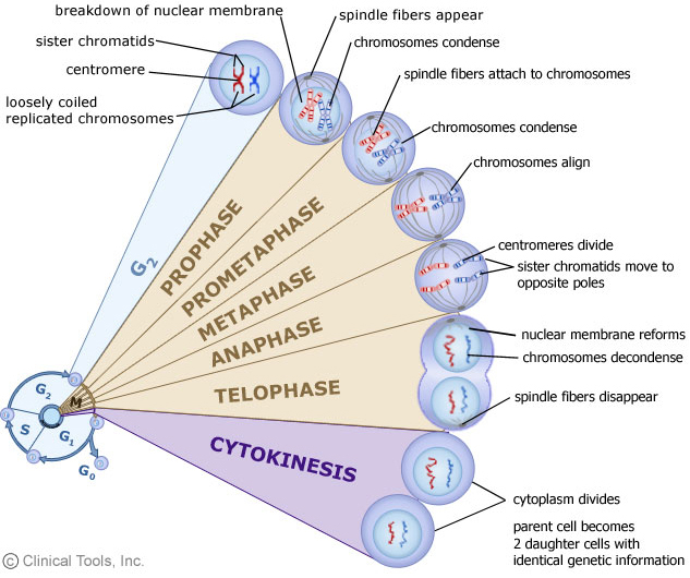
Interphase: 分裂间期; Prophase: 分裂前期; Metaphase: 分裂中期; Anaphase: 分裂后期; Telophase: 分裂末期

• Histone: 组蛋白

• Nucleosome: 核小体

•Chromosome: 染色体; Chromatin: 染色质; eu- 真染色质; hetero- 异染色质

  • Sister chromotid 姐妹染色单体;
  • mitotic spindle 纺锤体
  • spindle microtubule纺锤丝

• Centromere: 中心粒; Telomere: 端粒

====================================================

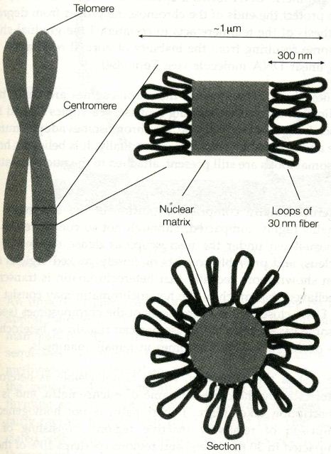
The familiar picture of a chromosome is actually that of the most highly condensed state at mitosis(which we reviewed in the blueprint of life [8]: eukaryotic structure of DNA 1(chromatin structure)).

As the daughter chromosomes are pulled apart by the mitotic spindle at cell division, the fragile centimeters-long chromosomal DNA would certainly be sheared by the forces generated, were it not in this highly compact state.


mitosisi chromosome

picture above is from Instant Notes in Molecular Biology

  • As we can see from the picture, the chromosomal loops fan out from a central scaffold or nuclear matrix region consisting of protein(which we talked about last section). One possibility is that consecutive loops may trace a helical path along the length of the chromosome.
  • The centromere is the constricted region where the two sister chromatids are joined in the metaphase chromosome. This is the site of assembly of the kinetochore, a protein complex which attaches to the microtubules of the mitotic spindle.

(The microtubules act to separate the chromotids at anaphase)

The DNA of the centromere has been shown in yeast to consist merely of a short AT-rich sequence of 88 bp, flanked by two very short conserved regions, although in mammalian cells, centromeres seem to consist of rather longer sequences, and are flanked by a large quantity of repeated DNA, known as satellite DNA.

  •  Telomeres are specialized DNA sequence that form the ends of the linear DNA molecules of the eukaryotic chromosomes. A telomoere consists of up to hundreds of copies of a short repeated sequence(5′-TTAGGG-3′ in humans), which is synthesized by the enzyme telomerase in a mechanism independent of normal DNA replication.

The telomeric DNA forms a special secondary structure, the function of which is to protect the ends of the chromosome proper from degradation.(Independent synthesis of the telomere acts to counteract the gradual shortening of the chromosome resulting from the inability of normal replication to copy the very end of a linear DNA molecule—we will talk about this when reaching DNA replication)


Interphase chromosome

In interphase(S phase, to be exact), the genes on the chromosomes are being transcribed and DNA replication takes place. During this time, the chromosmes adopt a much more diffuse structure and cannot be visualized individually. It is believed, however, that the chromosomal loops are still present, attached to the nuclear matrix.

The blueprint of life [9]: eukaryotic structure of DNA 2(nucleosome)

 
Eukaryotic chromosome is packaged in hierarchical levels, mediated by various proteins.
DNA duplexNucleosome → Chromatin →Chromosome

CHROMOSOME STRUCTURE (source)

We talked about DNA duplex and chromatin; now it’s time to meet the basic unit of chromatin structure–nucleosome.
  • —Definition: nucleosome is the chromatin subunit that consists of DNAand a set of eight histone core proteins(complex of (H2A)2(H2B)2(H3)2(H4)2, →octamerMore loosely with one molecule of H1 )
Comparison

HISTONES, shown by prof. Dong

The proteins protect the DNA from the action of micrococcal nuclease.
Micrococcal Nuclease is an endoexonuclease that preferentially digests single-stranded nucleic acids The enzyme is also active against double-stranded DNA and RNA and all sequences will be ultimately cleaved.(wikipedia)
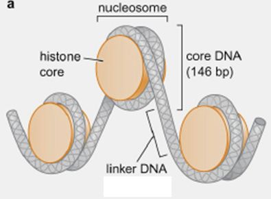
Digestion with nuclease leads to the loss of H1, yielding a very resistant structure consisting of 146 bp of DNA associated very tightly with the histone octamer.  The structure, known as the nucleosome core, is structurally very similar whatever the source of the chromatin.
  • —Adjacent nucleosome is connected by a varied length (10-100 bp, average 55bp)of DNA, called “linker DNA

LINKER DNA, shown by prof. Dong

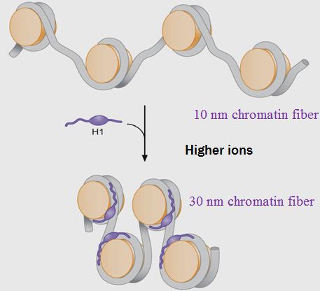
  • One molecule of linker histone H1 binds to the linker DNA between nucleosome.

H1 HISTONE, image from harmonybihar.org

H1 also acts to stabilize the point at which the DNA enters and leaves the nulceosome core.

H1 HISTONE, shown by prof. Dong

↑—Chromatins at different packaging levels
—30 nm chromatin fiber: condensed form
—10 nm chromatin fiber : less-condensed form, like a thread of beads
——————————————————————————-
Summary of Nucleosome Structure:

NUCLEOSOME STRUCTURE, image from bricker.tcnj.edu

——————————————————————————-
Higher Order Structure
The organization of chromatin at the highest level seems rather similar to that of prokaryotic DNA(see THE BLUEPRINT OF LIFE [7]: PROKARYOTIC CHROMOSOME STRUCTURE OF DNA). Even the size of the loops is approximately the same, up to aorund 100 kb of DNA, although there are many more loops in a eukaryotic chromosome.
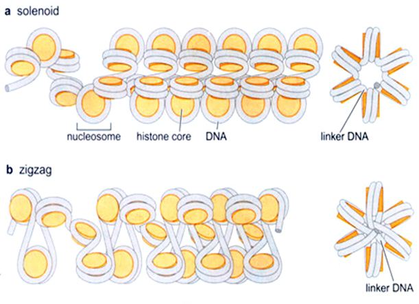
The loops are constrained by interaction with a protein complex known as the nuclear matrix. The DNA in the loops is in the form of 30 nm fiber, and the loops form an array about 300 nm across.
—”Solenoid model “of 30-nm chromatin fiber
—6 nucleosomes per turn
—“Zigzag model” of 30-nm chromatin fiber
—6 nucleosomes per turn, longer linker DNA may be required

HIGHER ORDER STRUCTURE, shown by prof. Dong

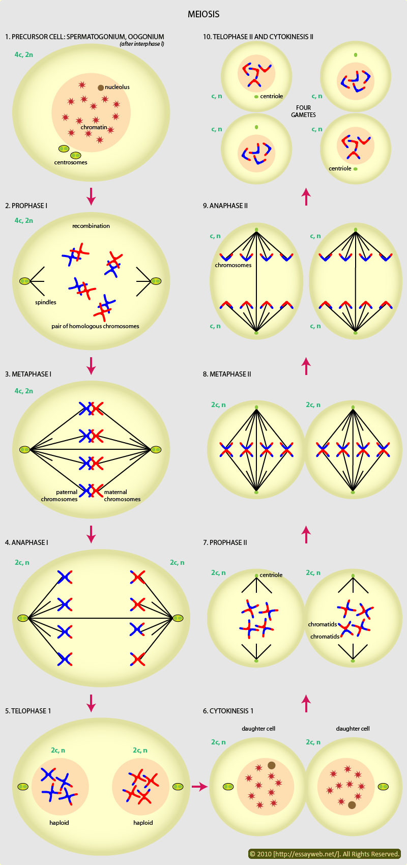
Genetics [9] Reproduction 2: Meiosis

During the process of reducing the number of chromosomes by half, the combination of alleles are rearranged to give recombinant gametes. Two distinct processes are involved. These are independent assortment of chromosomes and crossing-over.

Meiosis involves twosuccessive divisions, resulting in producing four cells, each containing half the number of chromosomes of the mother cell. There is NO replication between the two divisions.

We call the two divisions meiosis I and meiosis II, as in Chinese 减数第一次分裂 and 减数第二次分裂.  Here we will introduce more of meiosis than meets in high school textbooks.


Meiosis I

meiosis I is divided into prophase, metaphase, anaphase and telophase. Although they have the same names as the four phases in typical mitosis, behavior of chromosomes in these four phases in meiosis is very different from that in mitosis.

In prophase each chromosome pairs with its homolog (copy of the same chromosome inherited from the other parent). The paired chromosomes are called bivalents.  Each pair is held together by chiasmata(plural of chiasma). The exchange of genetic material is known as crossing-over.

chiasma (plural: chiasmata), in genetics, is thought to be the point where two homologous non-sisterchromatids exchange genetic material during chromosomal crossover during meiosis (sister chromatids also form chiasmata between each other, but because their genetic material is identical, it does not cause any change in the resulting daughter cells). (from wikipedia)

  • Prophase of meiosis is subdivided into five stages: leptotene, zygotene, pachytene, diplotene, and diakinesis.
  • In leptotene(literal translation:thin threads), the chromatin is seen to condense into very long thin strands, that appear tangled in the nucleus. As prophase proceeds chromosomes become shorter and thicker.
  • At zygotene(literal translation: yoked/linked threads), homologous chromosomes are seen as partially paired structures. They are still very elongated at this stage and chromosome pairs may overlap or interwine.
  • By patchytene(thick threads), pairing is complete, though it still isn’t possible identify clearly the individual chromatids in each bivalent.
  • As the homologous chromosomes begin to separate, transition from patchytene to diplotene(double threads) occurs.

This process begins at the centromeres and the bivalents are seen to be held together by chiasma.

The nuclear membrane and the nucleolus breaks down.

The four chromatids in each bivalent become identifiable and individual chiasma clearly to be identified.

  • Chromosomes carrying on condensing, the cell moves into diakinesis(moving apart), the final subdivision of prophase I.

The scheme below shows the five stages in prophase in a simple way:


At metaphase I the nuclear envelope breaks down, the bivalents lie across the equator of the cell with their centromeres attached to the microtbules←similar to the spindle in mitosis.

The dynamic action of the spindle causes one member of each homologous cell to move to the opposite poles of the cell.

↑WHY wouldn’t the action of the spindle tear apart the sister chromatids?

BECAUSE at metaphase the sister chromatids are held together by proteins called cohesions, which holds chiasmata in place and so holds the chromosomes together.


At anaphase I the cohesions in the chromosome arms are cut, allowing the homologs to separate.

In telophase I, two neclei form around the segregating chromosomes and a degree of chromosome decondensation is observed.


Meiosis II

The process of meiosis II closely resembles that of the typical mitosis.

In prophase II the chromosomes are seen to recondense within the two nuclei. At metaphase II, the nuclear membrane breaks down and chromosoms rearranged at the equator , the centromere splits and…At anaphase II and telophase II,the initial dipliod cell has divided into four…we already knew these in high school.

What we didn’t know in high school is that each of the  four haploid cells has a different genotype. And in many instances the group of four haploid cells may remain together and is known as a tetrad.


SUMMARY OF MEIOSIS

MEIOSIS, image from www.picstopin.com

the blueprint of life [8]: eukaryotic structure of DNA 1(chromatin)

• Mitosis: 有丝分裂; Meiosis: 减数分裂

Interphase: 分裂间期; Prophase: 分裂前期; Metaphase: 分裂中期; Anaphase: 分裂后期; Telophase: 分裂末期

• Histone: 组蛋白 hhistidine 组氨酸

• Nucleosome: 核小体

•Chromosome: 染色体; Chromatin: 染色质; eu- 真染色质; hetero- 异染色质

===================================================

The total length of DNA in a eukaryotic cell depends on the species, but it can be thousands of times as much as in a prokaryotic genome.

Eukaryotic chromosome is made up of a number of discrete bodies called chromosomes. The DNA in each chromosome is believed to be a single linear molecule, which can be up to several centimeters long.

All these each contain a long linear DNA molecules, which must be packaged into the nucleus, a space of approximately the same volume  as a bacterial cell

SO, much longer DNA chains packaged into a space of the same volume as a bacterial cell? → for example, 2 cm of DNA length versus ~10 µm of cell size for fruit fly; most condensed form of human chromosome is about ~2 µm long = 10,000× packing ratio

the obvious result is in their most highly condensed forms, the chromosomes have an enormously high DNA concentration: perhaps 200 mg/ml.!

The feat of packing is accomplished by the formation of a highly organized complex of DNA and protein, known as the chromatin, a nucleoprotein complex.       (←our hero today, has finally showed up.)

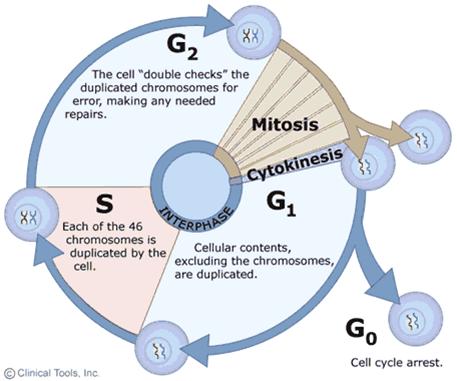
Chromosomes greatly alter their level of compectness as cells progress through the cell cycle, vary between highly condensed chromosomes at metaphase(just before the cell division), and very much more diffuse structures in interphase.(This implies the existence of different levels of organization of chromatin)

mitosis G1 S G2


mitotis phases


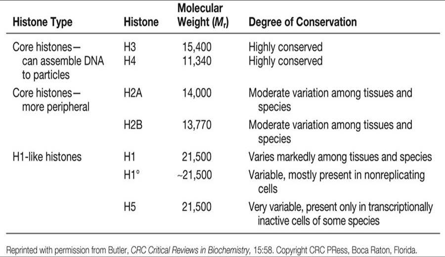
More than 50% of the mass of chromatin is protein. Most of the protein in eukaryotic chromatin consists of histones, of which there are five families: H2A, H2B, H3  and H4, known as the core histones, and H1.

The core histones are small proteins, with masses between 10 and 20 kDa, and H1 histones are a little larger at around 23 kDa.

The unified atomic mass unit (symbol: u) or dalton (symbol: Da) is the standard unit that is used for indicating mass on an atomic or molecular scale (atomic mass). One dalton is approximately the mass of a nucleon and is equivalent to 1 g/mol.[1] It is defined as one twelfth of the mass of an unbound neutral atom of carbon-12 in its nuclear and electronic ground state,[2] and has a value of 1.660538921(73)×10−27 kg.[3]

All histones proteins a large positive charge; between 20 and 30% of their sequences consist of the basic amino acids, lysine and arginine. This means that histones will bind very strongly to the negatively charged DNA in forming chromation.

amino acids in English?

Members of the same histone class(family) are very highly conserved between relatively unrelated species, for example between plants and animals, which testifies to their crucial role in  the chromation.

Within a given species, there are normally a number of closely similar variants of a particular class, which may be expressed in different tissues, and at different stages in development.

There is not much similarity in sequence between the different histone classes, but structural studies have shown that the classes so share a similar tertiary structure, suggesting that all hisotnes are ultimately evolutionarily related.

H1 histones are somewhat distinct from the other histone classes in a number of ways; in addition to their larger size, there is more variation in H1 sequences both between and within species than in other classes. Histone H1 is more easily extracted from bulk chromatin, and seems to be present in roughly half the quantity of the other classes, of which there are very similar amounts.

Next section we will cover the distinct role of histone Hi in chromatin structure.

Mendel’s Genetics [7]: handling problems

In college, while learning genetics, you may be faced with data obtained from F1 and F2 generations of the crosses. And you are required to be able to recognize ratios in order to decide how many genes are involved, and whether or not epistasis (which we talked about last section) is taking place.


Example(1) 

A cross between two  pure-breeding white-fruited tomato plants produced and F1 generation which all plants had purple fruit. In the subsequent F2 generation 160 plants were obtained; of these 94 had purple fuit, and the rest had white fruit.

As we know nothing about the genes controlling fruit color in tomato, we must first ask ourselves a question: “DOES THE DATA FIT ANY OF THE KNOWN MENDELIAN RATIOS?”

Since only two phenotypes are involved, it can’t be 9:3:3:1, or 9:3:4 or any mendelian ratios with more than 2 numbers included.

On examination of ratios with 2 phenotypes, 9:7 looks like a possible candidate, but 3:1 may also fit.

  • In this case, to decide which is the best to fit the data, we introduce a new approach to this  problem: the Chi-square test.
Chi-square(χw² test)=
sum[(obseved expected)ˆ2/expected]
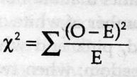
chi-square

(Xˆ2 is always calculated from original data, never from percentages, frequencies or proportions.)


  • If Xˆ2 is large, the data doesn’t fit. A perfect fit gives Xˆ2 a zero. BUT HOW LARGE IS LARGE?

In addition to the result of Xˆ2, we need another piece of info to determine “how large is large”. We need to know the degrees of freedom.

Degrees of freedom are one less than the number of classes. They tell us something about the number of independent numbers we have, which relates to the usefulness of our data.

In this example, we have two phenotypic classes, purple, and white. It means when we have counted the purple ones, the number of the white ones is fixed so we have only one degree of freedom.

If we had three classes, we would have two degrees of freedom: when the two classes have been counted, the number of the third is fixed.

As the degrees of freedom gets bigger, Xˆ2 gets bigger. So the answer of HOW LARGE IS LARGE depends on different degrees of freedom.

  • In this example, we determine which ratio is the best fit by comparing the value of Xˆ2

Observed result:                                   94 purple             66  white

Result predicted by 9:7 ratio        160×9/16 =90             160×7/16 =70

Xˆ2=[(94-90)ˆ2/90]+[(66-70)ˆ2/70]=0.41,with one degree of freedom

Result predicted by 3:1 ratio        160×3/4=120            160×1/4=40

Xˆ2=[(94-120)ˆ2/120]+[(66-40)ˆ2/40]=22.5,with one degree of freedom

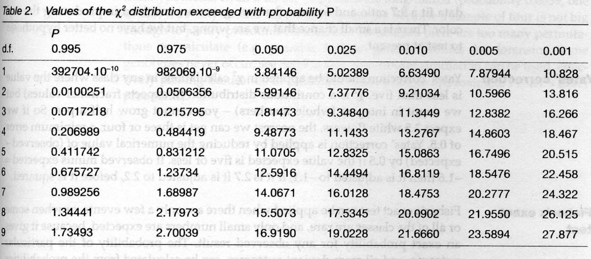
  • Using Xˆ2 probability tables can help quickly get to the value of Xˆ2.

the x2 ditribution table
X2 PROBABILITY TABLE, from “Instant Notes in Genetics”

How to use it?

Follow the line for the one degree of freedom(top line) to find the nearest values of xˆ2 above and below our value.

We can see that the value of 0.41 is between the probability of 0.975 and 0.050 with an affinity to 0.975, which means that if we repeated the experiment 1000 times, we have a probability close to 97.5% that the observed ratio would fit 9:7. The value of 22.5 is way beyond way exceeded with the probability of 0.001, which suggests that 3:1 ratio doesn’t fit the data. 

====================================================

Data analysis after class:

A cross between pure-breeding white-fruited and purple-fruited tomato plants produced and F1 generation which all plants had purple fruit. In the subsequent F2 generation 160 plants were obtained; of these 99 had purple fuit, 25 had red fruit, and 36 had white fruit.

====================================================

卡方检验是以χ2分布为基础的一种常用假设检验方法,它的无效假设H0是:观察频数与期望频数没有差别。

该检验的基本思想是:首先假设H0成立,基于此前提计算出χ2值,它表示观察值与理论值之间的偏离程度。根据χ2分布及自由度可以确定在H0假设成立的情况下获得当前统计量及更极端情况的概率P。如果P值很小,说明观察值与理论值偏离程度太大,应当拒绝无效假设,表示比较资料之间有显著差异;否则就不能拒绝无效假设,尚不能认为样本所代表的实际情况和理论假设有差别。


the blueprint of life [7]: prokaryotic chromosome structure of DNA

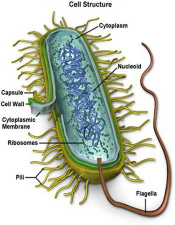
First, let’s make sure the anatomy of prokaryotes are familiar to us:

anayomy bacteria_cellProkaryotes are the simplest living cells, typically 1~10μm in diameter, and are found in all environmental niches from the guts of the animals to acidic hot springs.

  • They are bounded by a cell (plasma)membrane comprising a lipid bilayer, in which are embeded proteins that allow the exit and entry of small molecules.
  • Most prokaryotes also have a rigid cell wall outside the plasma membrane, which prevents the cell from swelling or shrinking in environments where osmolarity differs significantly from that inside the cell.
  • Sometimes the cell wall is surrounded by an (often) polysaccharide envelope called capsule.
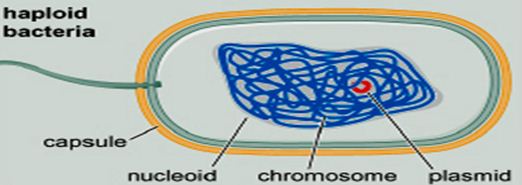
  • The cell interior (cytoplasm or cytosol) usually contains a single, circular chromosome compacted into a nucleoid and attached to the membrane.
  • There are no distinct subcelluar organelles in prokaryotes as in eukaryotes(except for the ribosomes核糖体).
  • The surface of a prokaryote may carry pili, which allow the prokaryote to attach to other cells and surfaces. Some prokaryotes also carry flagella, whose rotating motion allows the cell to swim.

====================================================


To talk about prokaryotic chromosome structure, we use E. coli (大肠杆菌)as the example.

cartoon e coli


  • —E. coli chromosome contains a single supercoiled circular DNA molecule of length 4.6 million bp.
  • —E. coli chromosome is highly folded: 1500 µm of DNA length versus ~1 µm of cell size, forming a structure called the nucleoid.
  • The nucleoid has a very high DNA concentration, perhaps 30~50 mg/ml, as well as containing all the proteins associated with DNA, such as polyerases, repressors(a protein that is determined by a regulatory gene, binds to a genetic operator, and inhibits the initiation of transcription of messenger RNA), etc.

nucleoid——————————————————————————-

DNA domains (loops)

DNA packingRemember this  famous electron micrograph of an E. coli cell we showed before? The cell was carefully lysed, all the proteins removed and then, it was spread on an EM grid to  reveal all of its DNA.
Several of such experiments revealed one level of organization of the nucleoid.
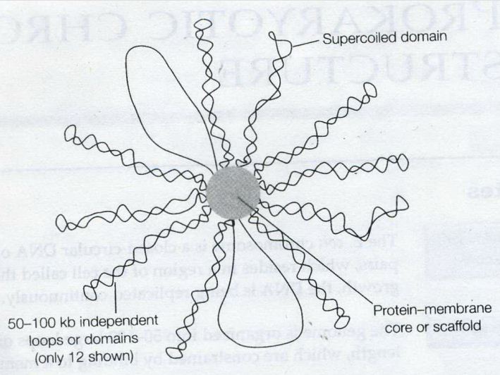
  • The DNA consists of 50~100 domains or loops, about 50~100 kb in size (kb: kilobase, a unit of measure of the length of a nucleic-acid chain that equals one thousand base pairs).
  • The ends of the loops are constrained by binding to a structure which probably consists of proteins attached to part of the cell membrane.
  • Not known whether the loops are static or dynamic, but one model suggests that the DNA may spool(wind) through sites of polyerase or other enzymic action at the base of the loops.

E coli DNA instant notes

 image above is from”Instant Notes in Molecular Biology”

Supercoiling of the genome

The E. coli chromosome as a whole is negatively supercoiled, although there is some evidence that indicates individual domains may be supercoiled independently. Electron micrographs indicate that some domains may not be supercoiled, perhaps because the DNA has become broken in one strand, where other domains clearly do contain supercoils.

The domains may be topologically independent. There is, however, no real biochemical evidence for major differences in the level of supercoiling in different regions of the chromosome in vivo


DNA-binding proteins

The looped DNA domains  of the chromosome are constrained further by inter-action with a number of DNA-binding proteins.

The most abundant of these are protein HU, a small basic (+charged) dimeric protein, which binds DNA non-specifically by the wrapping of the DNA aorund the protein.

And H-NS (formerly known as the protein H1), a monomeric neutral protein, which also binds DNA non-specifically in terms of sequence, but seems to have a preference for regions of DNA which are intrinsically bent.

These proteins are sometimes known as histone-like proteins, and have the effect of compacting the DNA, which is essential for the packaging of the DNA into the nucleoid, and of stabilizing and constraining the supercoiling of the chromosome.

the blueprint of life [6]: Chromosomal Structure of DNA 1


                                                       vocabulary

•Nucleus: 细胞核; Nucleolus: 核仁; Nucleoid: 类核

• Mitosis: 有丝分裂; Meiosis: 减数分裂

Interphase: 分裂间期; Prophase: 分裂前期; Metaphase: 分裂中期; Anaphase: 分裂后期; Telophase: 分裂末期

• Histone: 组蛋白

• Nucleosome: 核小体

•Chromosome: 染色体; Chromatin: 染色质; eu- 真染色质; hetero- 异染色质

• Centromere: 中心粒; Telomere: 端粒

====================================================

  • —WHAT is chromosome:
   Structure containing the genes of a cell and made of a single DNA molecule and its associated proteins.

CHROMOSOMES OF EUKARYOTES, shown by prof. Dong

CHROMOSOMES OF E. COLI, shown by prof. Dong

  • HOW long is DNA in an chromosome

    how long is DNA in a chromosome
    HOW LONG IS DNA, shown by prof. Dong.

→A chromosome is too long to fit into a cell without compaction.

  • WHY is DNA packed into chromosomes
  1. Chromosome is a compact form of the DNA that readily fits inside the cell
  2. To protect DNA from damage
  3. DNA in a chromosome can be transmitted efficiently to both daughter cells during cell division
  4. Chromosome confers an overall organization to each molecule of DNA, which regulates gene expression as well as recombination
 ——————————————————————————
Diversity of chromosomes

in terms of:

  • Shape: circular or linear
  • Number: species-specific

eg. -fruitfly 8; -human 46; -horse 64; -dog 78; -chicken 78

-maize 20; -rice 24; -wheat 42

  • Copy number: haploid单倍体, diploid双倍体, polyploid 多倍体
  • Overall structure: highly different between prokaryotes 原核生物and eukaryotes真核生物
——————————————————————————-
Next section we will first talk about prokaryotic chromosome structure of DNA.