the blueprint of life [11]: DNA replication 1

DNA replication

Definition: the process of copying a parental DNA molecule to form two daughter DNA molecules.

  • Introduction to DNA replication
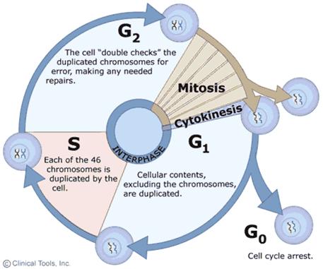
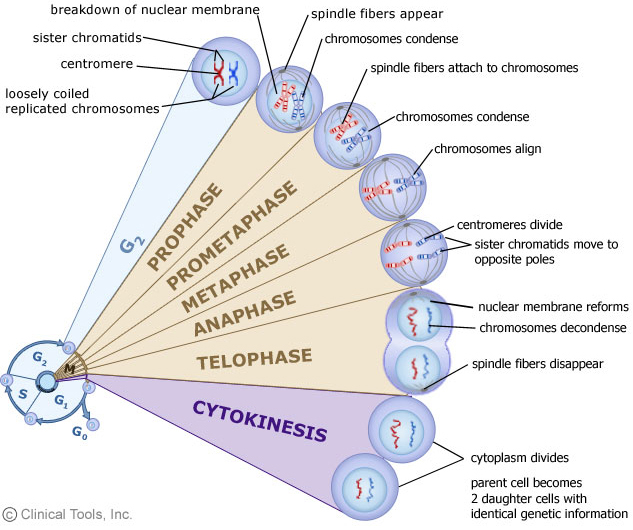
  1. DNA replication is essential for cell proliferation, i.e. mitosis, meiosis.
  2. DNA replication is a complex endeavor involving a series of enzyme activities.→see “DNA polymerases”
  3. DNA replication is performed in a semiconservative and semidiscontinuous mode.→see “DNA replication is semi-conservative”
  4. —DNA replication has 3 stages: initiation, elongation and termination.→next section
  5. —DNA replication is tightly regulated, involving various protein-protein, protein-DNA interactions.
  6. —DNA replication of prokaryote and eukaryote shares similar features, but is distinctive in details.
  •  Chemical Reaction of DNA replication

Essentials

1. Substrate: deoxynucleoside triphosphates(dNTPs)
2. Template: a primer-template junction
       DNA is synthesized by extending the 3’ end of the primer (free 3’-OH is required)
       – RNA primer or priming from a nick in DNA
3. Enzymes: DNA polymerases etc
4. Energy supply: Hydrolysis of pyrophosphate (PPi) is the driving force for DNA synthesis
5. Ions involved: Mg++ or Zn++
  • DNA polymerases
  • DNA polymerase I—
    • Pol I was the first enzyme discovered with polymerase activity, and it is also the best characterized one.
    • —Although abundant in cells (400/cell), Pol I is NOT the primary enzyme involved with bacterial DNA replication.
    • Main functions of Pol I: — (1) Fill any gaps in the new DNA that result from the removal of the RNA primer by its 5’ -3’ polymerase activity;   (2) Remove a new mispaired base by proofreading (校读)3’-5’ exonuclease (外切酶) activity. (3)Remove the RNA primer by its 5’-3’ exonuclease activity
The 3′–>5′ exonuclease activity intrinsic to several DNA polymerases plays a primary role in genetic stability; it acts as a first line of defense in correcting DNA polymerase errors. A mismatched basepair at the primer terminus is the preferred substrate for the exonuclease activity over a correct basepair. (source)
  • DNA polymerase III
    • The primary polymerase in DNA replication, although lower in abundance (15/cell)than pol  →referred to as “replicase
    • functions: (1) 5’→3’ polymerase activity; (2) 3’→5’ exonuclease activity – proofreading
    • Catalytic efficiency: much higher than pol I→High processivity and polymerization rate
    • A multi-unit complex: “holoenzyme” (全酶)
  • DNA replication is semi-conservative 

Bet you all have learned it in high school, and the famous experiment by Meselson and Stahl. We still need to go over the points again as they are essentially important for what we will learn next.

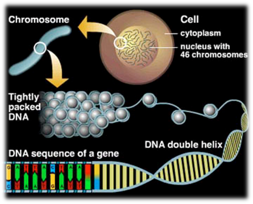
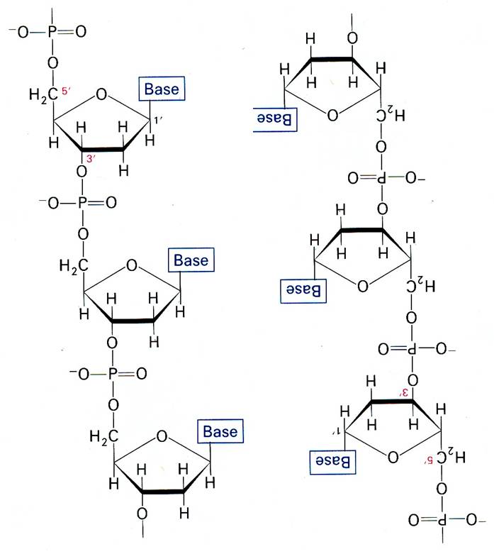
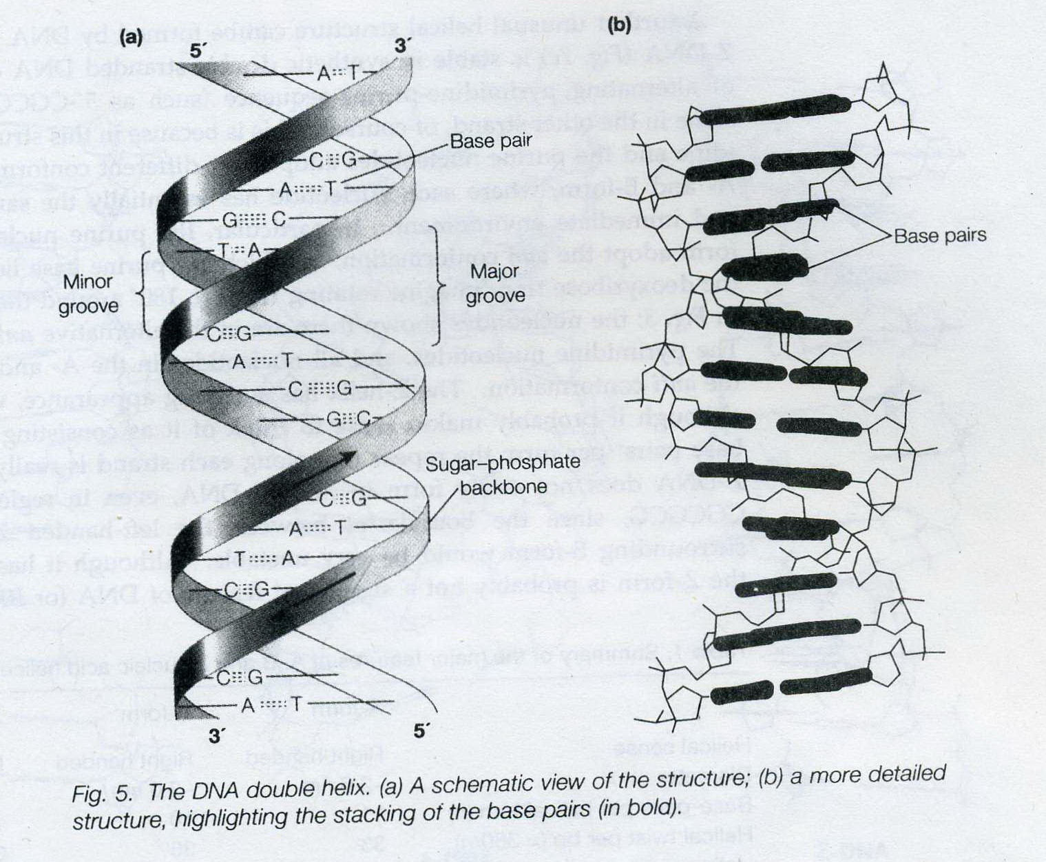
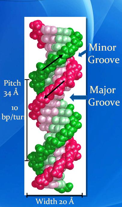
The key to the mechanism of DNA replication is the fact that each strand of the DNA double helix carries the same information-their base sequences are complementary (we talked about this in THE BLUEPRINT OF LIFE [2]: PRIMARY AND SECONDARY STRUCTURE OF DNA).

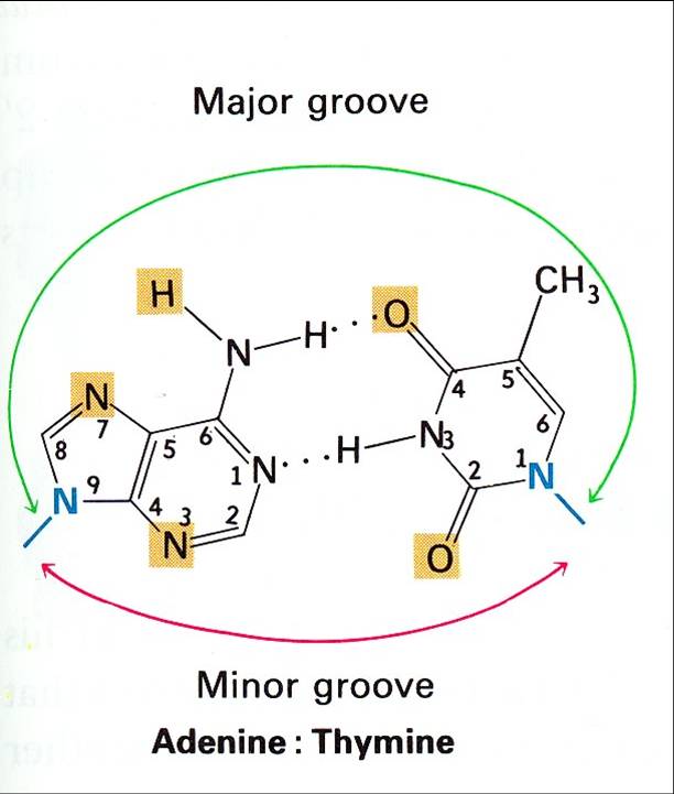
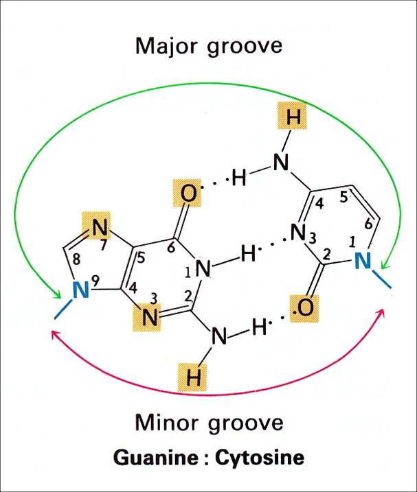
During replication, the two parental strands separate and each acts as a template (that’s right, the template for DNA replication is DNA itself!)to direct the enzyme-catalyzed synthesis of a new complementary daughter strand with the normal base-pairing rules (A-T, C-G)

This semi-conservative mechanism was demonstrated experimentally in 1958 by Meselson and Stahl.

Hypotheses:

In the experiment:

E. coli cells were grown for several generations in presence of the stable heavy isotope 15N so that their DNA became fully density labeled (both strands are 15N labeled: 15N/15N)

The cells were then transferred to medium containing only normal 14N and, after each cell division, DNA was prepared from a sample of the cells and analyzed on a CsCI gradient using the technique of equilibrium (isopycnic) density gradient centrifugation, which separates molecules according to differences in buoyant density.

After the first cell division, when the DNA had replicated once, it was all of hybrid density, in a position on the gradient half way between fully labled (15N /15N )and fully light (14N/14N). After the second generation in 14N, half of the DNA was hybrid density and half fully light.

Thus, two of the hypotheses were denied, left us with the semi-conservative mechanism.

After each subsequent generation, the proportion of 14N/14N increased, while some DNA of hybrid density persisted. Thus the semi-conservative mode of DNA replication is confirmed: each daughter molecule contains one parental strand and one newly-synthesized strand.

DNA replication & PCR, General Biology, Open Courses at UC-Berkeley

General Biology, Great open courses given by Gary L. Firestone, Michael Meighan Jasper D. Rine and Jennifer A. Doudna, professors at UC-Berkeley.

There is more to DNA replication than we talked about yesterday so I tried to upload this video to help you learn more. 

“tried? ”

“Well, turned out ‘ DNA replication and PCR open course at Berkeley.mp4 exceeds the maximum upload size (8 MB)for this site.'”

So I would just provide the link where you can watch it online.

After watching it, maybe you will find more than just DNA replication: you may as well find how it feels going to college.

I look forward to it every time I have finished an open course online. Hope you do, too.

>>click here to watch the video


the blueprint of life [8]: eukaryotic structure of DNA 1(chromatin)

• Mitosis: 有丝分裂; Meiosis: 减数分裂

Interphase: 分裂间期; Prophase: 分裂前期; Metaphase: 分裂中期; Anaphase: 分裂后期; Telophase: 分裂末期

• Histone: 组蛋白 hhistidine 组氨酸

• Nucleosome: 核小体

•Chromosome: 染色体; Chromatin: 染色质; eu- 真染色质; hetero- 异染色质

===================================================

The total length of DNA in a eukaryotic cell depends on the species, but it can be thousands of times as much as in a prokaryotic genome.

Eukaryotic chromosome is made up of a number of discrete bodies called chromosomes. The DNA in each chromosome is believed to be a single linear molecule, which can be up to several centimeters long.

All these each contain a long linear DNA molecules, which must be packaged into the nucleus, a space of approximately the same volume  as a bacterial cell

SO, much longer DNA chains packaged into a space of the same volume as a bacterial cell? → for example, 2 cm of DNA length versus ~10 µm of cell size for fruit fly; most condensed form of human chromosome is about ~2 µm long = 10,000× packing ratio

the obvious result is in their most highly condensed forms, the chromosomes have an enormously high DNA concentration: perhaps 200 mg/ml.!

The feat of packing is accomplished by the formation of a highly organized complex of DNA and protein, known as the chromatin, a nucleoprotein complex.       (←our hero today, has finally showed up.)

Chromosomes greatly alter their level of compectness as cells progress through the cell cycle, vary between highly condensed chromosomes at metaphase(just before the cell division), and very much more diffuse structures in interphase.(This implies the existence of different levels of organization of chromatin)

mitosis G1 S G2


mitotis phases


More than 50% of the mass of chromatin is protein. Most of the protein in eukaryotic chromatin consists of histones, of which there are five families: H2A, H2B, H3  and H4, known as the core histones, and H1.

The core histones are small proteins, with masses between 10 and 20 kDa, and H1 histones are a little larger at around 23 kDa.

The unified atomic mass unit (symbol: u) or dalton (symbol: Da) is the standard unit that is used for indicating mass on an atomic or molecular scale (atomic mass). One dalton is approximately the mass of a nucleon and is equivalent to 1 g/mol.[1] It is defined as one twelfth of the mass of an unbound neutral atom of carbon-12 in its nuclear and electronic ground state,[2] and has a value of 1.660538921(73)×10−27 kg.[3]

All histones proteins a large positive charge; between 20 and 30% of their sequences consist of the basic amino acids, lysine and arginine. This means that histones will bind very strongly to the negatively charged DNA in forming chromation.

amino acids in English?

Members of the same histone class(family) are very highly conserved between relatively unrelated species, for example between plants and animals, which testifies to their crucial role in  the chromation.

Within a given species, there are normally a number of closely similar variants of a particular class, which may be expressed in different tissues, and at different stages in development.

There is not much similarity in sequence between the different histone classes, but structural studies have shown that the classes so share a similar tertiary structure, suggesting that all hisotnes are ultimately evolutionarily related.

H1 histones are somewhat distinct from the other histone classes in a number of ways; in addition to their larger size, there is more variation in H1 sequences both between and within species than in other classes. Histone H1 is more easily extracted from bulk chromatin, and seems to be present in roughly half the quantity of the other classes, of which there are very similar amounts.

Next section we will cover the distinct role of histone Hi in chromatin structure.

The blueprint of life [5]: spectroscopic and thermal properties of DNA

UV absorption
  • DNA absorbs UV light due to the conjugated aromatic nature of the bases; the sugar-phosphate backbone does not contribute appreciably(perceptibly/measurably) to absorption.

The wavelength of maximum absorption of light by both DNA and RNA is 260 nm, which is conveniently distinct from the λmax of protein(280 nm).

The absorption properties of DNA can be used for detection, quantitation and assessment of property.

  • Hypochromicity

UV absorption at 260nm is greatest for isolated nucleotides, intermediate for single-stranded DNA(ssDNA) or RNA, and least for double-stranded DNA(dsDNA)

The classical term for the change in absorbance is hypochromicity. For example, dsDNA is hypochromic (from the Greek for ‘less colored‘) relative to ss DNA, which is hypochromic relative to isolated nucleotides.

—Thermodynamics of DNA
  •  —Denaturation:  the transition of macromolecule from the native state to the denatured state. For DNA, under denaturing conditions (heating or high pH), double helix is separated to generate single-stranded form.
—Melting Temperature(Tm):  the temperature at which the rise in A260(absorbance at 260nm) is half complete during denaturation.
—Factors that affect the Tm
1. G+C content: the higher G+C content, the higher Tm.
2.— Ionic strength: The Tm increases as the cation (+) concentration              increases. like Na+, K+ or Mg2+.
3.— High pH or Agents that disrupt H-bonds or interfere with base                 stacking: formamide (甲酰胺)or urea (尿素)will decrease the Tm.
4. —The imperfect hybridization between related but not completely            complementary strands will reduce the Tm, about 1 °C for each                percent mismatch.
The process of denaturation can be observed conveniently by the increase in absorbance as double-stranded nucleic acids are converted to single strands
  • —Renaturation: Process of a macromolecule returning to its native 3-D structure. For DNA this involves the two strands of denatured DNA basepairing to restore the nature form of dsDNA. Also known as reannealing (重退火).
—Requirements for renaturation:
—1. Proper salt concentration: neutralize electrostatic repulsion
—2. Proper reannealing temperature: 20-25℃ below Tm
—Determinants for renaturation efficiency
(DNA renatures on cooling, but will form fully double-stranded native DNA only if the cooling is sufficiently slow to allow the complementary strands to anneal.)
1. Extent of base matching and
2. copy of matching regions
Thus, repetitive DNA renatures faster than single copy DNA

RENATURATION CURVES FOR E.COLI &MOUSE DNA, shown by prof. Dong


The blueprint of life [4]: Tertiary Structure of DNA

Finally, we come to the last part of the molecular structure of DNA.

4. Tertiary Structure of DNA – superhelix structure

—Advanced folding and intertwining of DNA molecules over the secondary structure .

DNA topology
 1. —Linear DNA:
  • —Commonly seen in eukaryotes,
  • with extreme length,
  • complementary sequence
  • included in the chromatin (the combination or complex of DNA and proteins that make up the contents of the nucleus of a cell)
  • interacting with other cellular components.
2. —Circular DNA:
(DNA frequently occurs in nature as closed-circular molecules, where the two single strands are each circular and linked together. The number of links is known as the linking number(Lk).)
  • Usually seen in prokaryotes, e.g. plasmid (质粒), circular bacterial chromosomes and many viral DNA molecules
  • —cccDNA (covalently closed circular DNA) → supercoiled or Relaxed;         ncDNA (nicked circular DNA): a nick (缺刻)formed by breaking  one phosphodiester bond—
  • two complementary single strands are each joined into circles, 5′ to 3′, and are twisted around one another by the helical path of DNA.
  • The molecule has no free ends and the two single strands are linked together a number of times corresponding to the number of double-helical turns in the molecule.

DNA topology1 DNA topology2

cccDNA  Topology 

  •  Supercoiled DNA

        Negative superhelix: natural status of cccDNA with less intra-               molecular tension (underwound effect)

        Positive superhelix: unnatural status with higher tension                           (overwound effect)

  • Relaxed circular DNA: intermediate between negative superhelix and positive superhelix
  • —Topological Equation of cccDNA
       L   =  T   +   W
—
—L=Linking number=total number of times one strand of the double helix links the other
—T=Twisting number= the number of times one strand completely wraps around the other strand
—W=Writhing number= the number of times that the long axis of the double helical DNA crosses over itself in 3-D space
    —   Features
—      1. The linking number of a closed-circular DNA is a topological                      property, that is one which cannot be changed without breaking            one or both of the DNA backbones. (A molecule of a given                            linking number is known as a topoisomer. Topoisomers differ                    only in their linking number)
        2. —Twisting number
              For B DNA, T>0(10 bp per turn)
              For A DNA, T>0(10.5 bp per turn)
              For Z DNA, T<0 (12 bp per turn)
        3. —Writhing number
             Relaxed: W=0
             Negative supercoils: W<0
             Positive supercoils: W >0

TWIST &WRITHE, from Instant Notes in Molecular Biology(3rd edition)

  •  Topoisomerases (enzymes used to regulate the level of supercoiling of DNA molecules)

topoisomerase
TOPOISOMERASE FUNCTIONING, shown by prof.Dong

To alter the linking number of DNA, the enzymes must transiently break one or both stands. There are two classes of topoisomerases:
Type I  Topoisomerase nick one strand of the DNA, changing 1                                  Linking-number at a time(+/-1 Lk)
Type II Topoisomerase, which requires the hydroloysis of ATP, break                   two strands of DNA, changing 2 Linking-number at a                                       time(+/-2Lk); also able to unlink DNA molecules.
Most topoisomerases reduce the level of positive or negative supercoiling, that is, they operate in the energetically favorable direction. (However, DNA gyrase, a bacterial type II enzyme, uses the energy of ATP hydrolysis to introduce negative supercoiling into hence removing positive supercoiling generated during replication.)
Topoisomerases are essential enzymes in all organisms; they are involved in replication, recombination and transcription.
Both type I and II enzymes are the target of anti-tumor agents in humans.
DNA superhelix
—Biological significance of Superhelix:
  • —DNA packing:
Eg.
DNA packing
This is a famous electron micrograph of an E. coli cell that has been carefully lysed, then all the proteins were removed, and it was spread on an EM grid to  reveal all of its DNA.
  • — DNA functioning:
        Negative supercoils serve as a store of free energy that aids in processes requiring strand separation, such as DNA replication and transcription; Strand separation can be accomplished more easily in negatively supercoiled DNA than in relaxed DNA.
1. DNA in cells is negatively supercoiled;
2.(-)supercoiling introduced by a topoisomerase II (gyrase 促旋酶) in prokaryotes and by nucleosome (核小体)in eukaryotes
3.Cruciform or bubble structures introduced by (-) supercoiling are potential protein-binding sites

The blueprint of life [3] secondary and some special structures of DNA

Let’s pick up where we dropped, the secondary structure of DNA.

IN FACT, a number of different forms of nucleic acid double-helix have been observed and studied, all having the basic pettern of two helically-wound antiparallel strands.
Polymorphism (多样性)of DNA  Secondary Structure
―due to conformational changes of sugar-ring on the nucleotide chain.
1. B-form:
  • right-handed
  • the structure identified by Watson and Crick,
  • the most common form,
  • believed to be the idealized form of the structure adopted by virtually all DNAin vivo (in the living body of a plant or animal), or,  at physiological (characteristic of or appropriate to an organism’s healthy or normal functioning) pH and salt concentration.

characterized by:

  • a helical repeat of 10bp/turn (although now it is known that ‘real’ B-DNA has a repeat closer to 10.5bp/turn);
  • the presence of base pairs lying on the helix axis and almost perpendicular to it;
  • having well-defined, deep major and minor grooves.
2. A-form: 
  • right handed
  • adopted by DNA in vivo under unusual circumstances, (conversed from B-form in low moisture (<75%))
  • presents in certain DNA-protein complexes

characterized by:

  • a helical repeat of 11 bp/turn.
  • the presence of base pairs tilted with respect to the helix axis, and actually lying off the axis.
  • being the helix formed by RNA and DNA-RNA hybrids. (Similar to some RNA-DNA duplex or RNA-RNA duplex.)
3. Z-form:
  • left-handed
  • formed by stretches of alternating pyrimidine-purine sequence, e.g. GCGCGC, especially in negatively supercoiled DNA in high saline (盐) solution.
  • not easily form even in DNA regions of repeating GCGCGC, since the boundaries between the left-handed Z-form and the surrounding B-form would be very unstale

characterized by:

  • a zigzag (锯齿型) pattern where its name comes from
  • 12 bp/turn

(1)

A. B, Z-DNA
slide shown by prof.Dong,COMPARISON AMONG A-,B-,Z-DNA

(2)
A.B.Z-DNA instant notes
picture from Instant Notes in Molecular Biology (3rd Edtion)
====================================================
Some special structures of DNA 
1. Inverted repeats and direct repeats
Inverted Repeat is functionally important as recognition sites on the DNA for the binding of a variety of proteins (e.g. restriction and modification enzymes)
Inverted Repeat is either discontinuous or continuous,
Continuous → palindromic structure (回文结构:inverted repetitions of base sequence over the two strands form a symmetric structure )
Discontinuous→ hairpin & cruciform (发卡、十字结构:self complementary within each of the strands )

palindromic structure

hairpin structure & cruciform structure
hairpin structure & cruciform structur

2. DNA triplex (The intramolecular triplex (H-DNA) as an example)

shown by prof.Dong, DNA TRIPLEX

• AG-rich strand vs. CT-rich strand with Hoogsten hydrogen bond
(a quick glance at Hoodsten hydrogen bond:

image from wikipedia/hoogsten base pair

• Mirror inverted repeat
• a barrier for different DNA and RNA polymerases, and so it is a negative regulator for gene expression.
3. G-quadruplex structure
G-rich sequence(s) to form 4-stranded structure by unusual G-G
H-bonds

shown by prof.Dong, G-AUADRUPLEX

the blueprint of life [2]: primary and secondary structure of DNA

Molecular structure of DNA:

professor Dong first showed us where DNA is:

molecular structure of DNA
slide shown by Prof.Dong,WHERE IS DNA?

Then he introduced the molecular structure of DNA with 4 parts:

1. Primary structure of DNA:

Arrangement of nucleotides along a DNA chain.

Annie: So…the primary structure of DNA is a line.

Conventionally, the repeating monomers of DNA are represented by their single letters A, T, G, C.

Professor: There’s a convention to write the DNA sequence with 5′ at the left, that is, in a 5′ to 3′ orientation from left to right.

====================================================

2. Secondary structure of DNA—stabilized partial structure formed by polymers of nucleotide

Professor Dong: First, please familiarize yourselves with Chargaff’s Rule:

A+G=T+C & G+T=A+C

↓↓

A=T & G=C

(Chargaff’s Rule)

(Based on analysis of the chemical composition of duplex DNA in the early 1950s, E. Chargaff deduced these rules about the amounts of different nucleotides in DNA.)

Professor: The secondary structure of DNA is a partial structure formed by polymers of nucleotide. The structure is referred to as the double-helix structure.

Two separate chains of DNA are wound around each other, each following a helical (coiling) path, resulting in a right-handed double helix structure.

In 1953, Watson and Click proposed the DNA double-helix structure based on Chargaff’s Rule and DNA Crystallography and X-ray diffraction images of DNA structure by Wilkins and Franklin.  (Rosalind Franklin, who was not that well-known as Watson, Click and Wilkins but apparently played a equally significant role in the discovery of the structure.The whole was later nominated Nobel Prize but her, is that even fair?)

—————————————————————————

The backbone of duplex DNA is a serious of phosphodiester group (the covalent linkage of a phosphate group between the 5′-hydroxyl of one sugar and the 3′-hydroxyl of the next, that is , repeats of P-sugar unit) linked by phosphodiester bond.

—————————————————————————
The strands are joined noncovalently by hydrogen bonding between the bases on opposite strands, to form base pairs.
There are around 10 base pairs per turn in the DNA double-helix. The two strands are oriented in opposite directions in terms of their 5’to3′ direction(the nucleotides in one strand is opposite to their direction in the other strand).
stand direction
More crucially, the two strands are complementary in terms of sequence. The bases hydrogen-bond to each other as purine-pyrimidine pairs which have very similar geometry and dimensions.
         A–T:  2 H-bonds ;    C–G:  3 H-bonds
   5’- A   T   G    T   C -3’
   ¦¦    ¦¦  ¦¦¦   ¦¦   ¦¦¦
   3’- T   A   C   A   G -5’

 Thus, the sequence of one strand uniquely specifies the sequence of the other, with all that which implies for the mechanism of replication of DNA and its transcription to RNA.

——————————————————————————-
Professor Dong added:
Between the backbone stands run the major and minor grooves.

In a detailed analysis of DNA structure, there are two types of grooves that can be seen; the major groove has the nitrogen and oxygen atoms of the base pairs pointing inward toward the helical axis, while in the minor groove,the nitrogen and oxygen atoms point outwards;

major groove A_T
Shown by prof.Dong, MAJOR GROOVE A-T

major groove GC
Shown by prof.Dong, MAJOR GROOVE G-C

—Major Groove                        —Minor Groove
—Depth: 8.5 Å                             —Depth: 7.5 Å
—Width: 11.7 Å                          —Width: 5.7 Å
Å
Definition: Symbol for Ångström, a unit equal to 0.1 nanometer, mainly used in expressing sizes of atoms, lengths of chemical bonds, and wavelengths of electromagnetic radiation.
Supplement: The unit is named after the Swedish physicist, Ångström, Anders Jonas.
instant notes

picture from Instant Notes in Molecular Biology

Professor: The major groove is more dependent on base composition. and major grooves and minor grooves are also recognition and binding sites for certain  protein factors, and are involved in the regulation of gene expression.

——————————————————————————

grooves
slide shown by Prof.Dong

Professor: Summary of “Double Helix” Model (B-DNA):

  • —Right Handed Double Helix
  • —Outside: P-Sugar backbone
  • Inside: Base pairing linked by H-bonds
  • —Minor and Major grooves
 (note: bp=base pair(s))

the blueprint of life[1]

Of course the blueprint of life is DNA.

DNA is important because it is the genetic material ←contain all the info for the synthesis and functioning of a living form duplicate and passes through to the next generation.

Hearing this Annie questioned herself: DNA is not the genetic for ALL viruses. RNA “blueprints” for the rest of the viruses. Virus, though not even having a cell structure, is a form of life. So why not be fair, and say the blueprint of life are DNA and RNA?

Professor Dong continued,

Proofs:

  1. Bacterial Transformation Experiment

—Griffith, 1928

Professor: So what is the transforming principle? 

Annie: It could be a cool type of enzyme that moved the toxic part of S strain onto R strain…

Professor: Well, Enzyme did help a lot in the experiment we’ll talk about later, but it is not the hero of the story.

Annie: So what’s the story?

Professor:

—Avery et al., 1944

slide shown by Prof. Dong ,AVERY REPEATED GRIFFITH’S EXPERIMENT WITH MODIFICATION

Only DNA is responsible for the transformation.

Annie thought: Well, there still existed possibilities that DNA and some other things that were not sugar, lipid or protein cooperated to complete the transformation… The “other things” also contributed to the transformation but could not complete it without DNA?  In this case DNA is not the only one that is responsible,  

  1. T2 Bacteriophage Infection (Blender Experiment)

—Hershey &Chase, 1952

Experiment 1

Experiment 2

Radioactive labeling of proteins and DNA

The professor continued. So now let’s go deeper and see the chemical composition of DNA.

A 5-carbon sugar , hand in hand with base group and phosphorous group, thought Annie.

It’s not that simple as you learned in high school, said the professor. We need to know the chemical composition of the base group, deoxyribon and phosphorous group as well.

sugar group
slide shown by Prof. Dong,SUGAR GROUP

 Professor:

Base
slide shown by Prof. Dong,BASE GROUPS

Annie thought: Pyrimid-ine, Could it have anything to do with the Pyramid in Egypt?

The professor was explaining the structure and Annie didn’t interrupt him with her question.

After class, Annie searched the Internet for an answer, and this is what she found.

 The professor continued.

Bonds

P group——phosphester bond–Sugar—–glycosidic bond—–B group