同翅目 HOMOPTERA
| Order Hemiptera | |
| suborder Heteroptera * | Bugs |
| suborder Homoptera * | Cicadas, Hoppers, Psyllids, Whiteflies, Aphids, and Scale insects |
Most people tend to call anything with lots of legs a “bug.” However, to an entomologist, a “bug” is one of the 35,000 or so species of the order Hemiptera.
Hemiptera means “half wing” and refers to the fact that part of the first pair of wings is toughened and hard, while the rest of the first pair and the second pair are membranous. Hemipterans also have modified piercing and sucking mouthparts; some suck plant juices and are plant pests, while others can bite painfully.
A possibly paraphyletic group of insects known as the Homoptera is sometimes included within the Hemiptera, even though they lack the toughened areas on the first pair of wings.
Some entomologists group both Hemiptera and Homoptera within the group Heteroptera; others use the name Heteroptera for what we have called the Hemiptera and use Hemiptera for the Heteroptera.
Confused? So are we. Anyway, the Homoptera have the dubious distinction of being probably the most destructive insects of all. They include aphids, leafhoppers, cicadas, and scale insects: 45,000 species in all.
↑Quoted from University of California Museum Paleontology>arthropoda>uniramia>hemiptera
Hemiptera
Suborder Homoptera
Leafhoppers, Planthoppers, Treehoppers, Cicadas, Aphids, Psyllids, Whiteflies, Scale Insects
The name Homoptera, derived from the Greek “homo-“meaning uniform and “ptera” meaning wings, refers to the uniform texture of the front wings.
- Classification & Distribution
Hemimetabola
- incomplete development (egg, nymph, adult)
Hemipteroid
- closely related to Thysanoptera and Psocoptera
Distribution: Abundant worldwide. All species are terrestrial herbivores.
|
North America
|
Worldwide | |
|---|---|---|
| Number of Families |
38
|
60
|
| Number of Species |
6359
|
>32,000
|
- Physical Features
 |
Adults:
Immatures:
|
Major Families
- Cicadidae (Cicadas) — Nymphs live underground where they feed on the roots of trees and shrubs. Adults are the largest members of the Homoptera. Males produce loud songs to attract a mate.
- Cicadellidae (Leafhoppers) — This is the largest family of Homoptera and includes many pests of cultivated plants. Leafhoppers are important carriers of plant diseases — especially mycoplasmas.
- Membracidae (Treehoppers) — Ecologically similar to leafhoppers, these insects have a large pronotum that extends over most of the body. They often resemble thorns or small twigs.
- Cercopidae (Spittlebugs or Froghoppers) — Nymphs live on plant stems and produce a frothy defensive secretion around themselves. Adults are similar to leafhoppers in size and general appearance.
- Fulgoridae (Planthoppers) — This is one of eleven families classified as planthoppers (superfamily Fulgoroidea). These insects are ecologically similar to leafhoppers and treehoppers. Many species are oddly shaped and cryptically colored.
- Psyllidae (Psyllids or Jumping Plant Lice) — Small, aphid-like insects with 3-segmented beaks and 10 segmented antennae. Many species are covered with a woolly layer of wax.
- Aleyrodidae (Whiteflies) — Body and wings of adults are covered with a white powdery wax. Nymphs attach to the undersides of leaves and become immobile, resembling scale insects.
- Aphididae (Aphids, Plantlice) — Second largest family in the suborder Homptera. Many of these insects are pests of cultivated plants. Aphids are considered the most important carriers of viral plant diseases.
- Coccidae (Soft Scale insects) — This is one of 17 families that make up the superfamily Coccoidea (scale insects and mealybugs). Most species are sedentary during most of their life cycle and secreate a protective covering over their bodies. These insects are among the most common pests of cultivated plants.
====================================================↑Quoted from the General Entomology course at North Carolina State University >Resource Library > Compendium > Homoptera (© 2009 by John R. Meyer; Last Updated: 8 April 2009)
>Learn more about homoptera (www.insectsexplained.com)
====================================================
蝉科 Cicadidae
1. 草蝉 Mogannia sp.

2. 绿草蝉 Mogannia hebes

3. 蒙古寒蝉 Meimuna mongocica

4. 螗蝉Tanna japonensis

5. 鸣鸣蝉 Oncotympana maculaticollis


6. 蟪蛄Platypleura kaempferi

叶蝉科 Cicadellidae
1. 丽叶蝉 Calodia sp.


2. 华凹大叶蝉 Bothrogonia sinica

3. 条大叶蝉Atkinsoniella

4. 显脉叶蝉Paramesus sp.

5. 槽胫叶蝉Drabescus sp.

6. 窗耳叶蝉Ledra auditura

7. 隐纹大叶蝉Tettigoniella thalia

8. 横脊叶蝉Evacanthus sp.

9.白边拟大叶蝉Ishidaella albomarginata


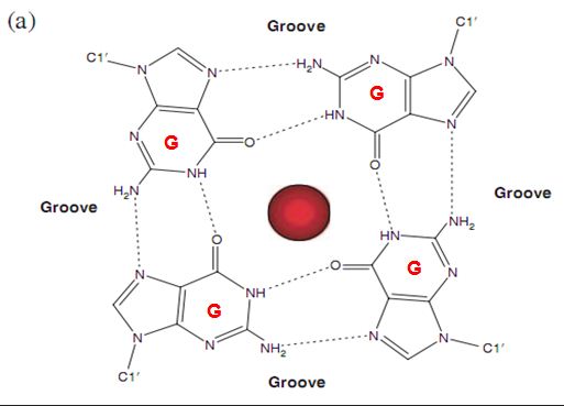
![The blueprint of life [4]: Tertiary Structure of DNA](https://apbiology.cn/wp-content/uploads/sites/8/2013/07/DNA-topology2.jpg)
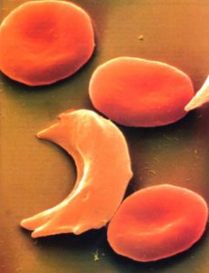
![Mendel’s Genetics [4]: examples of mutiple alleles](https://apbiology.cn/wp-content/uploads/sites/8/2013/12/genetics-4-blood-cell.jpg)
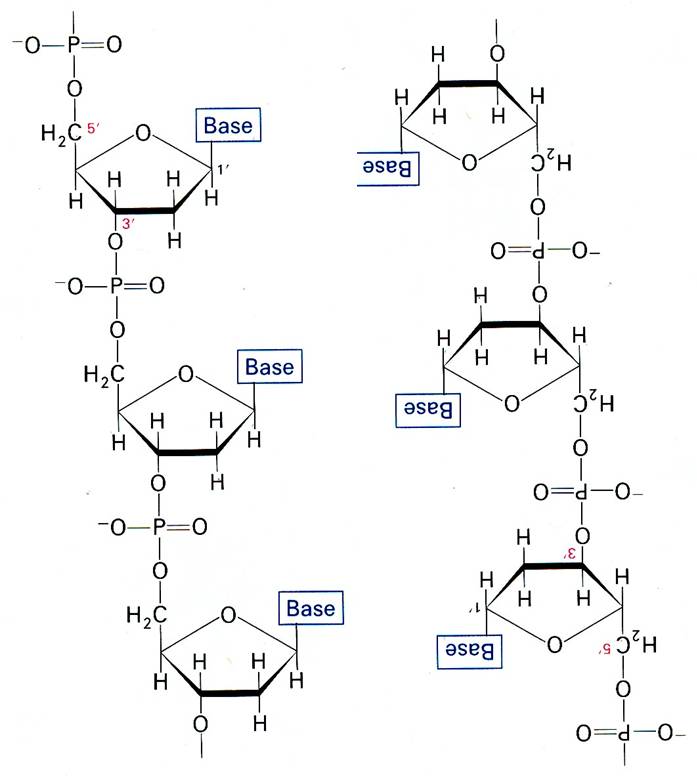
![The blueprint of life [3] secondary and some special structures of DNA](https://apbiology.cn/wp-content/uploads/sites/8/2013/07/dna2.jpg)
![Mendel’s Genetics[3]: Variations of the 3:1ratio](https://apbiology.cn/wp-content/uploads/sites/8/2013/12/genetics-3-snapdragons.jpg)
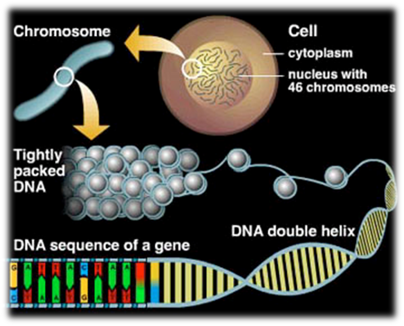


![the blueprint of life[1]](https://apbiology.cn/wp-content/uploads/sites/8/2013/06/Science-Human-Gene.jpg)