![the blueprint of life [10]: eukaryotic structure of DNA 3](https://apbiology.cn/wp-content/uploads/sites/8/2013/12/blueprint-8-eukaryotic-chromosome.jpg)
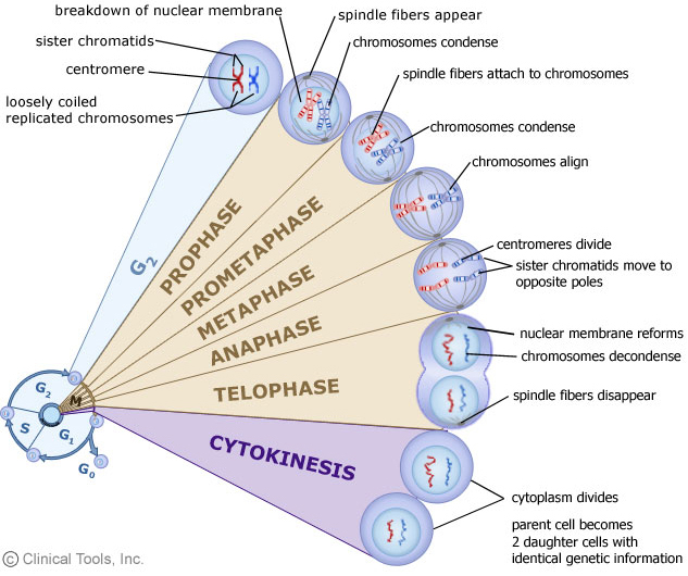
let’s go over the terms again. Make sure you all know what they mean.
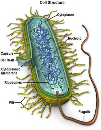
•Nucleus: 细胞核; Nucleolus: 核仁; Nucleoid: 类核
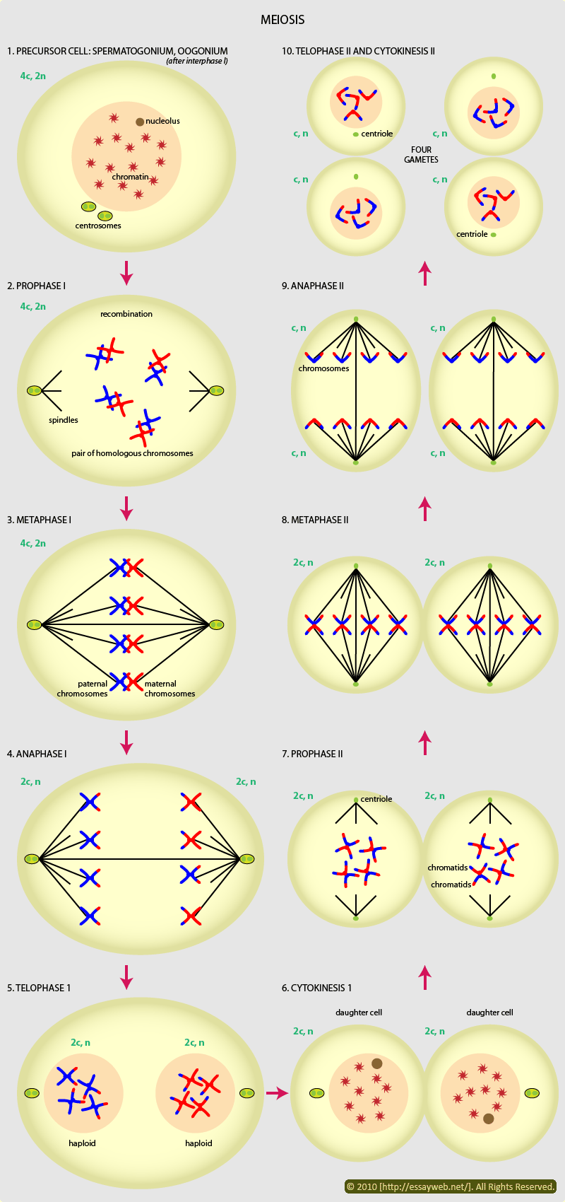
• Mitosis: 有丝分裂; Meiosis: 减数分裂
Interphase: 分裂间期; Prophase: 分裂前期; Metaphase: 分裂中期; Anaphase: 分裂后期; Telophase: 分裂末期
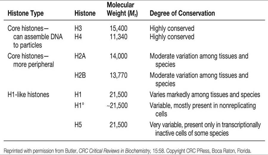
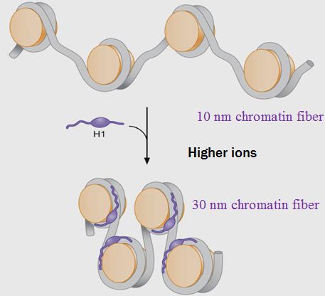
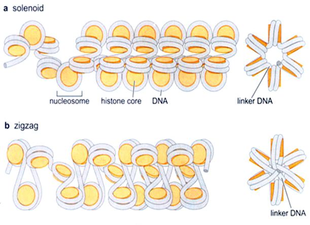
• Histone: 组蛋白
• Nucleosome: 核小体
•Chromosome: 染色体; Chromatin: 染色质; eu- 真染色质; hetero- 异染色质
- Sister chromotid 姐妹染色单体;
- mitotic spindle 纺锤体
- spindle microtubule纺锤丝
• Centromere: 中心粒; Telomere: 端粒
====================================================
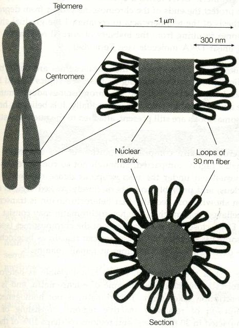
The familiar picture of a chromosome is actually that of the most highly condensed state at mitosis(which we reviewed in the blueprint of life [8]: eukaryotic structure of DNA 1(chromatin structure)).
As the daughter chromosomes are pulled apart by the mitotic spindle at cell division, the fragile centimeters-long chromosomal DNA would certainly be sheared by the forces generated, were it not in this highly compact state.
picture above is from Instant Notes in Molecular Biology
- As we can see from the picture, the chromosomal loops fan out from a central scaffold or nuclear matrix region consisting of protein(which we talked about last section). One possibility is that consecutive loops may trace a helical path along the length of the chromosome.
- The centromere is the constricted region where the two sister chromatids are joined in the metaphase chromosome. This is the site of assembly of the kinetochore, a protein complex which attaches to the microtubules of the mitotic spindle.
(The microtubules act to separate the chromotids at anaphase)
The DNA of the centromere has been shown in yeast to consist merely of a short AT-rich sequence of 88 bp, flanked by two very short conserved regions, although in mammalian cells, centromeres seem to consist of rather longer sequences, and are flanked by a large quantity of repeated DNA, known as satellite DNA.
- Telomeres are specialized DNA sequence that form the ends of the linear DNA molecules of the eukaryotic chromosomes. A telomoere consists of up to hundreds of copies of a short repeated sequence(5′-TTAGGG-3′ in humans), which is synthesized by the enzyme telomerase in a mechanism independent of normal DNA replication.
The telomeric DNA forms a special secondary structure, the function of which is to protect the ends of the chromosome proper from degradation.(Independent synthesis of the telomere acts to counteract the gradual shortening of the chromosome resulting from the inability of normal replication to copy the very end of a linear DNA molecule—we will talk about this when reaching DNA replication)
Interphase chromosome
In interphase(S phase, to be exact), the genes on the chromosomes are being transcribed and DNA replication takes place. During this time, the chromosmes adopt a much more diffuse structure and cannot be visualized individually. It is believed, however, that the chromosomal loops are still present, attached to the nuclear matrix.


![Genetics [9] Reproduction 2: Meiosis](https://apbiology.cn/wp-content/uploads/sites/8/2013/11/genetics10-meiosis.gif)
![Genetics [8]:reproduction 1](https://apbiology.cn/wp-content/uploads/sites/8/2013/11/genetics-9-dinosuar.jpg)
![the blueprint of life [8]: eukaryotic structure of DNA 1(chromatin)](https://apbiology.cn/wp-content/uploads/sites/8/2014/01/m-bio-8-chromotin.png)


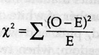
![Mendel’s Genetics [7]: handling problems](https://apbiology.cn/wp-content/uploads/sites/8/2013/12/chi-square.jpg)
 test)=
sum[(obseved expected)ˆ2/expected]
test)=
sum[(obseved expected)ˆ2/expected]
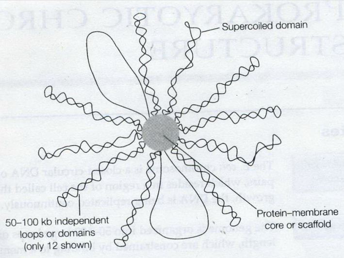
![the blueprint of life [7]: prokaryotic chromosome structure of DNA](https://apbiology.cn/wp-content/uploads/sites/8/2013/07/m-bio-7-bacteria.jpg)
 ——————————————————————————-
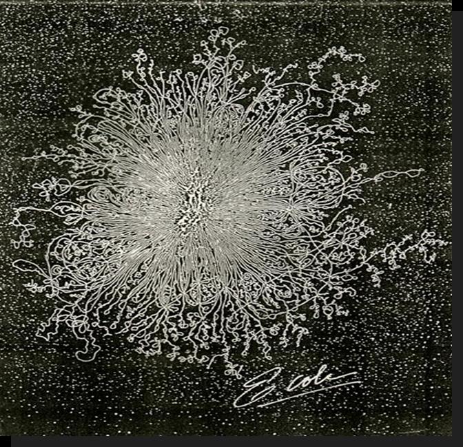 Remember this famous electron micrograph of an E. coli cell we showed before?