“Winter gives us the opportunity to stay inside and look outside. Snuggle up in thesofa, put a blanket over you,have a cup of hot cocoa, and enjoy the observations on this precious season.
“Winter is the time for comfort, for good food and warmth, for the touch of a friendly hand and for a talk beside the fire: It is the time for home. (the warmth in winter)
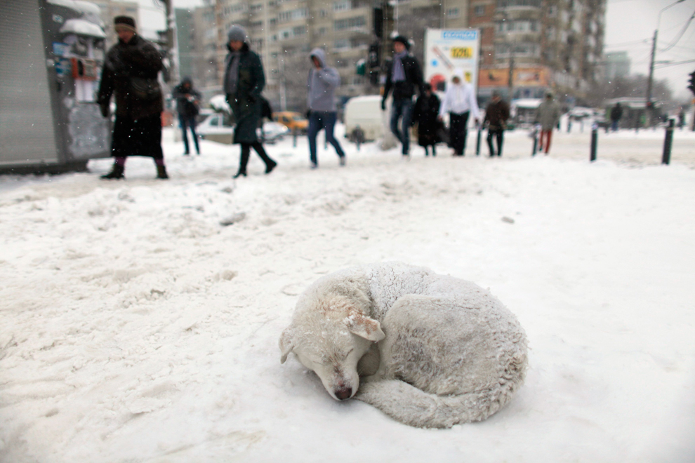
BUT what about those who don’t have a home? For them, winter is definitely not a time for comfort, but a time for freezing and hunger.
Homeless people could die of coldness, so could stray animals.
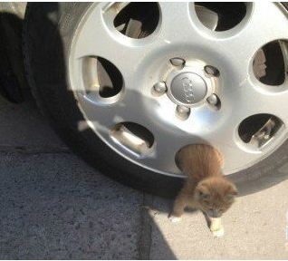
We see the homeless sleep in the subway stations or the underground, where it’s relatively warmer and free of the chilly wind. I don’t see many stray animals stay in the underground for night; perhaps they are afraid of humans. Most often they hide in trash cans, car engine compartment or inside the wheels or the tail pipes where it’s warmer, and to them, safer.
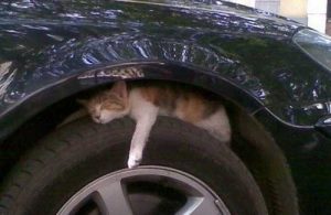
But the truth is it’s not safe at all sleeping inside any of the places above. Especially the latter three. In China every year countless cats are critically injured, even killed inside their “warm spots”. In the morning most people go downstairs, directly get in their cars, start the engine, and head for office. Chances are, a poor kitty was soundly sleeping under the hood of the car and didn’t get a chance to wake up and escape before the car moved and hurt her.
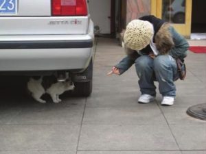
SO next time before starting your car, please take time to check inside the engine compartment, the wheels and the trail tube, see if any small animal is inside. Not just cats, rats, little yellow weasels, which are common here in Hefei are all possible.If you’ve checked and don’t see any of them but still aren’t sure, it’s also a very good idea to sound your horn for a few times and wait for seconds, as my mom always does. Hearing the noise, the small animals are alarmed and would come out and escape.
IF you forget to “release”the little thing and hurt it, please contact the veterinary hospital and get the animal medical treatment as soon as possible. Don’t try moving the animal if he/she is stuck in the wheel or the engine compartment. Don’t move your car, either.
Most of the homeless animals are homeless because some of us bought them and then abandoned them. Most of the wild animals would “break into” our city because we took their homes. Now they are just trying to survive in a strange place without sofa, fireplace or warm woods.Is there any reason why we shouldn’t at least give them a safer spot for overnight?
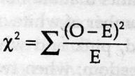
![Mendel’s Genetics [7]: handling problems](https://apbiology.cn/wp-content/uploads/sites/8/2013/12/chi-square.jpg)
 test)=
sum[(obseved expected)ˆ2/expected]
test)=
sum[(obseved expected)ˆ2/expected]
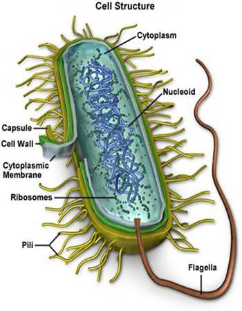
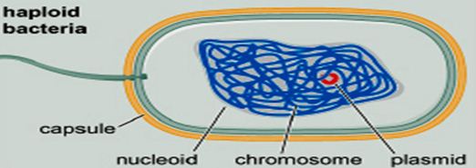
![the blueprint of life [7]: prokaryotic chromosome structure of DNA](https://apbiology.cn/wp-content/uploads/sites/8/2013/07/m-bio-7-bacteria.jpg)
 ——————————————————————————-
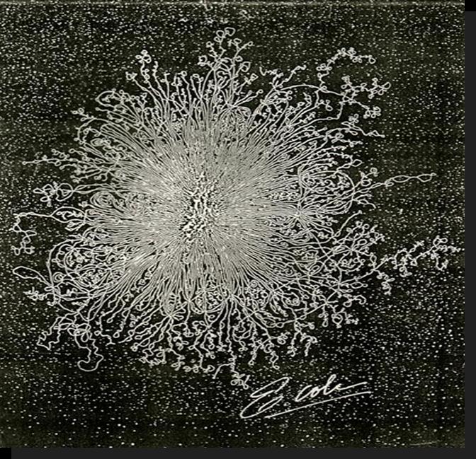 Remember this famous electron micrograph of an E. coli cell we showed before?
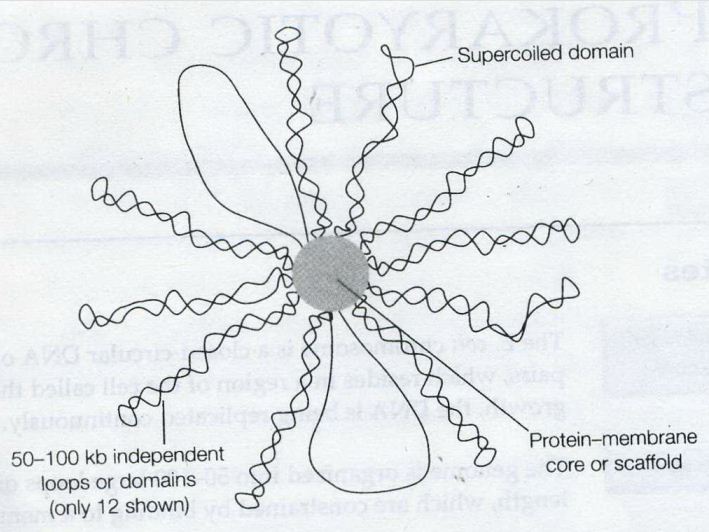


![the blueprint of life [6]: Chromosomal Structure of DNA 1](https://apbiology.cn/wp-content/uploads/sites/8/2013/12/m-bio-6-chromosomes.jpg)

 A novel you wouldn’t put down once opening the first page.


![Mendel’s Genetics [6]: Examples of epistasis](https://apbiology.cn/wp-content/uploads/sites/8/2013/12/genetics-3-snapdragons.jpg)
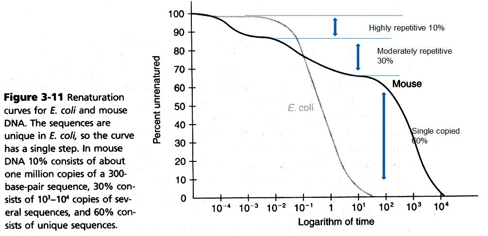
![The blueprint of life [5]: spectroscopic and thermal properties of DNA](https://apbiology.cn/wp-content/uploads/sites/8/2013/07/UV-absorption-of-DNA.jpg)