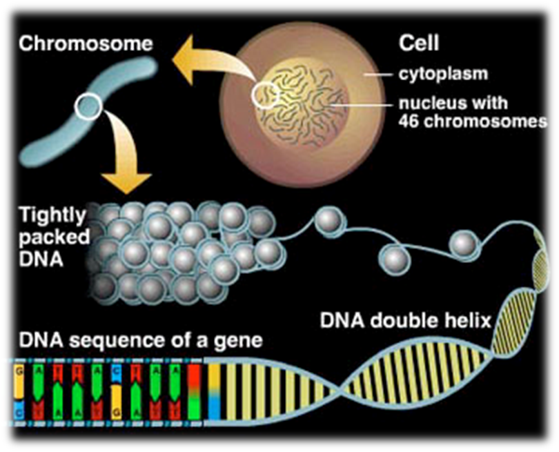
2. Secondary structure of DNA—stabilized partial structure formed by polymers of nucleotide
Professor Dong: First, please familiarize yourselves with Chargaff’s Rule:
A+G=T+C & G+T=A+C
↓↓
A=T & G=C
(Chargaff’s Rule)
(Based on analysis of the chemical composition of duplex DNA in the early 1950s, E. Chargaff deduced these rules about the amounts of different nucleotides in DNA.)
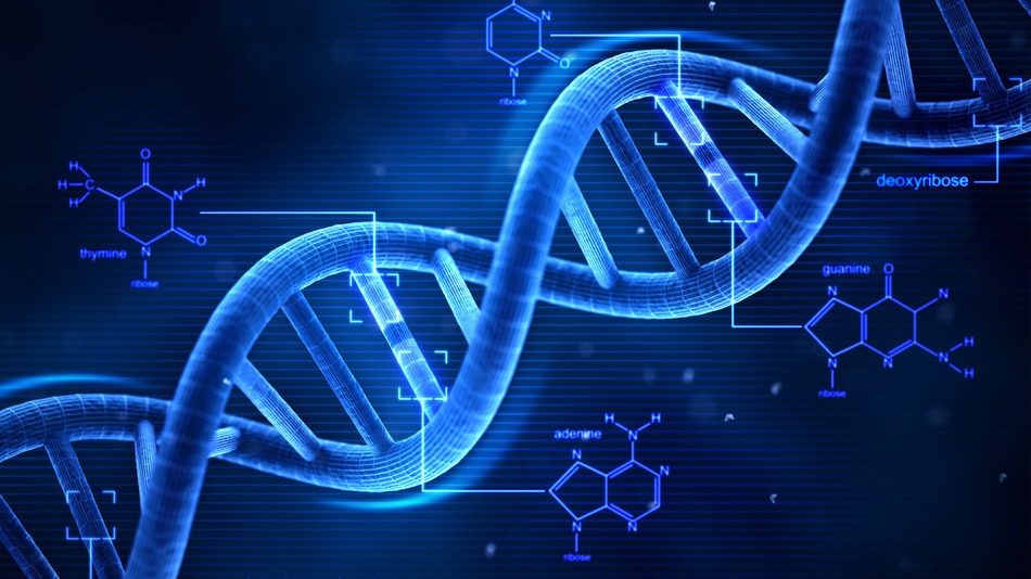
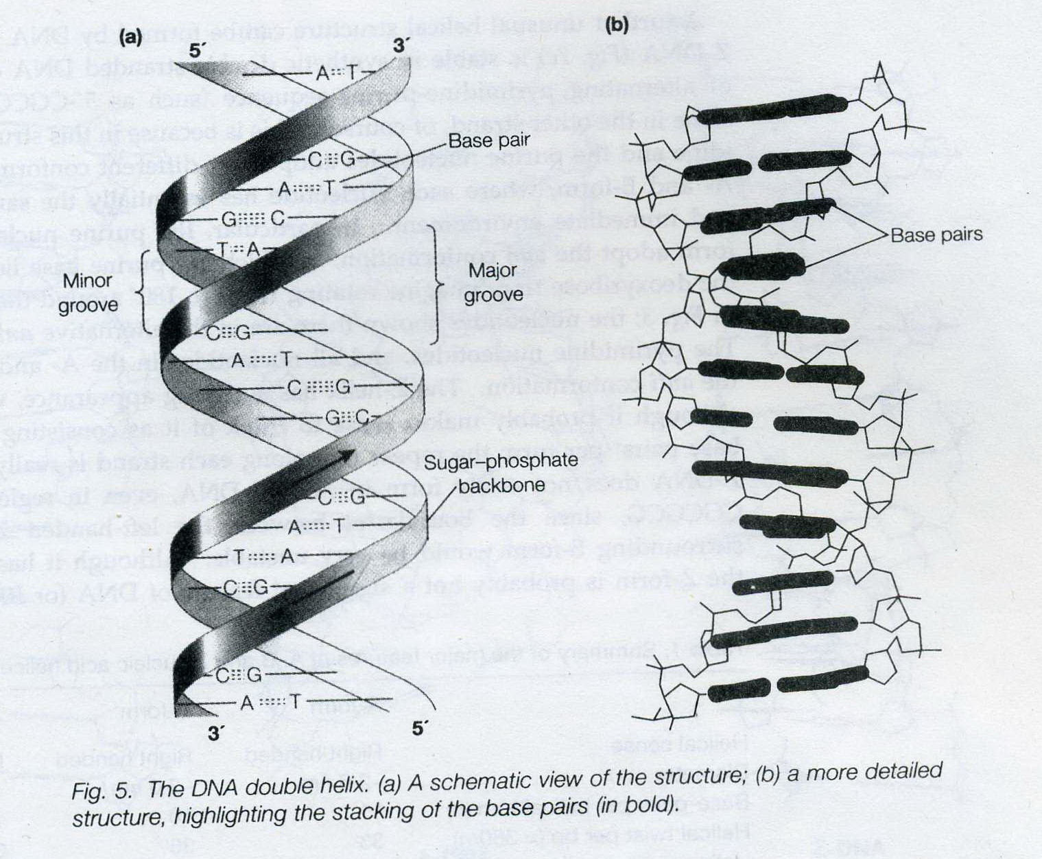
Professor: The secondary structure of DNA is a partial structure formed by polymers of nucleotide. The structure is referred to as the double-helix structure.
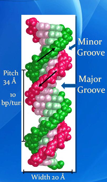
Two separate chains of DNA are wound around each other, each following a helical (coiling) path, resulting in a right-handed double helix structure.
In 1953, Watson and Click proposed the DNA double-helix structure based on Chargaff’s Rule and DNA Crystallography and X-ray diffraction images of DNA structure by Wilkins and Franklin. (Rosalind Franklin, who was not that well-known as Watson, Click and Wilkins but apparently played a equally significant role in the discovery of the structure.The whole was later nominated Nobel Prize but her, is that even fair?)
—————————————————————————
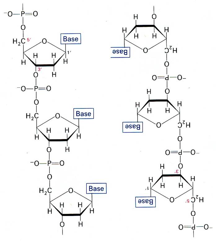
The backbone of duplex DNA is a serious of phosphodiester group (the covalent linkage of a phosphate group between the 5′-hydroxyl of one sugar and the 3′-hydroxyl of the next, that is , repeats of P-sugar unit) linked by phosphodiester bond.
—————————————————————————
The strands are joined noncovalently by hydrogen bonding between the bases on opposite strands, to form base pairs.
There are around 10 base pairs per turn in the DNA double-helix. The two strands are oriented in opposite directions in terms of their 5’to3′ direction(the nucleotides in one strand is opposite to their direction in the other strand).
More crucially, the two strands are complementary in terms of sequence. The bases hydrogen-bond to each other as purine-pyrimidine pairs which have very similar geometry and dimensions.
A–T: 2 H-bonds ; C–G: 3 H-bonds
5’- A T G T C -3’
¦¦ ¦¦ ¦¦¦ ¦¦ ¦¦¦
3’- T A C A G -5’
Thus, the sequence of one strand uniquely specifies the sequence of the other, with all that which implies for the mechanism of replication of DNA and its transcription to RNA.
——————————————————————————-
Professor Dong added:
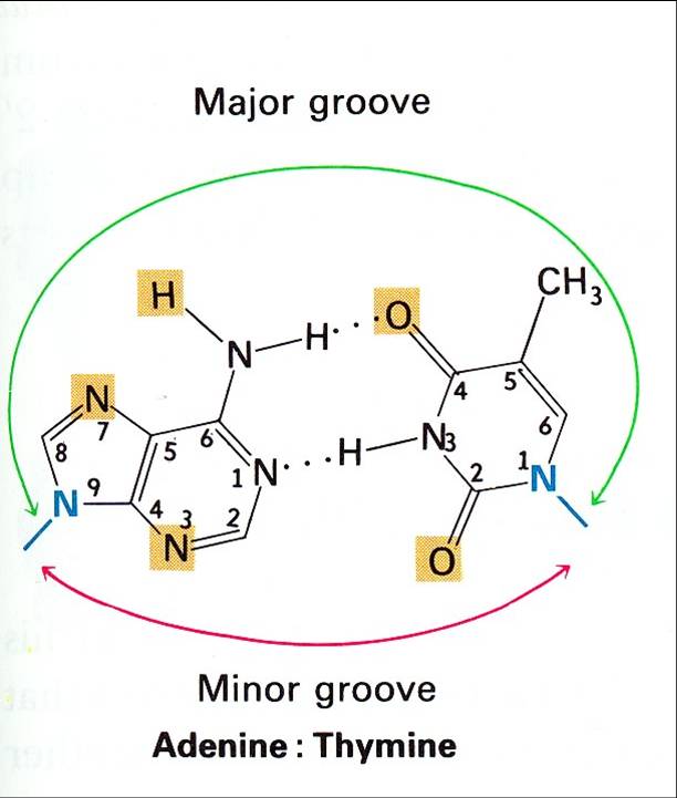
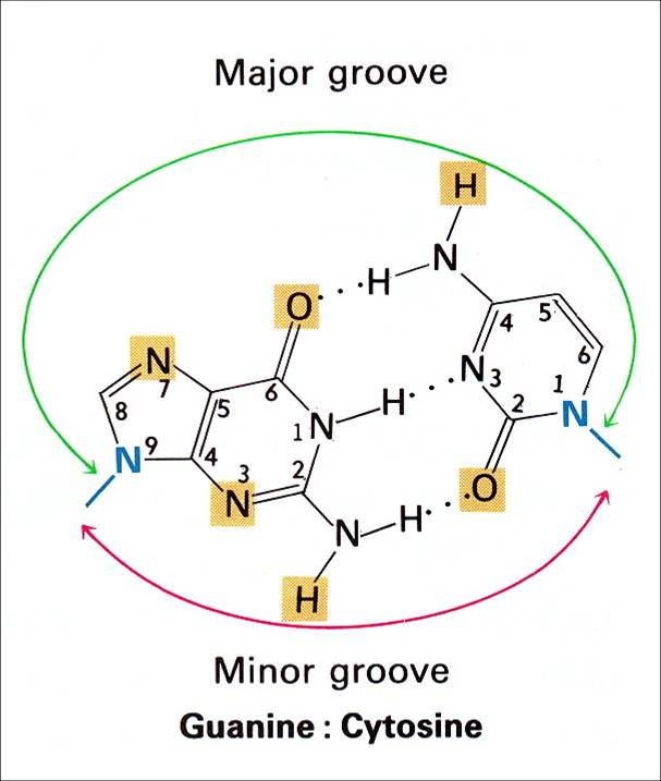
Between the backbone stands run the major and minor grooves.
In a detailed analysis of DNA structure, there are two types of grooves that can be seen; the major groove has the nitrogen and oxygen atoms of the base pairs pointing inward toward the helical axis, while in the minor groove,the nitrogen and oxygen atoms point outwards;
Shown by prof.Dong, MAJOR GROOVE A-TShown by prof.Dong, MAJOR GROOVE G-C
Major Groove Minor Groove
Depth: 8.5 Å Depth: 7.5 Å
Width: 11.7 Å Width: 5.7 Å
Å
Definition: Symbol for Ångström, a unit equal to 0.1 nanometer, mainly used in expressing sizes of atoms, lengths of chemical bonds, and wavelengths of electromagnetic radiation.
Professor: The major groove is more dependent on base composition. and major grooves and minor grooves are also recognition and binding sites for certain protein factors, and are involved in the regulation of gene expression.
——————————————————————————
slide shown by Prof.Dong
Professor: Summary of “Double Helix” Model (B-DNA):